Fig. 5.
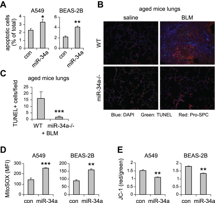
miR-34a promotes lung epithelial cell apoptosis and mitochondrial dysfunction. A: human lung epithelial cells A549 and BEAS-2B were transfected with 50 nM control (con) mimics or miR-34a mimics. Three days after transfection, cells were collected, and apoptosis determined by FITC-annexin V/propidium iodide assay. Values are means ± SD; n = 3. *P < 0.05 and **P < 0.01 compared with con miR group. B and C: aged WT and miR-34a−/− mice were intratracheally treated with BLM. Three weeks after instillation, mice were killed, and lung sections prepared. Apoptotic cells were assessed by terminal deoxynucleotidyl transferase dUTP nick-end labeling (TUNEL) assay (green fluorescence). B: slides were costained with pro-surfactant protein C (SPC; red). C: the number of TUNEL+ cells was counted (original magnification, × 20). ***P < 0.001 compared with WT+ group. D: A549 and BEAS-2B cells were transfected with 50 nM con mimics or miR-34a mimics. Three days after transfection, mitochondrial ROS were determined using MitoSOX Red followed by flow cytometry. E: A549 and BEAS-2B cells were transfected as in D. Three days after transfection, cells were collected, and mitochondrial membrane potential was determined by JC-1 assay. D and E: values are means ± SD; n = 3. **P < 0.01 and ***P < 0.001 compared with con miR group. MFI, mean fluorescence intensity.

