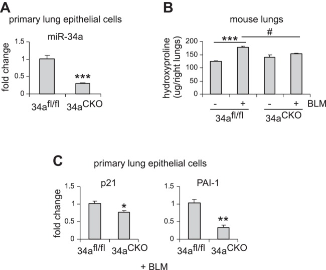
Fig. 6.
Alveolar epithelial ablation of miR-34a protects mice from experimental lung fibrosis. A: alveolar epithelial type 2 (ATII) conditional knockout mice (miR-34aCKO) and control miR-34afl/fl mice were administered tamoxifen (75 mg/kg body wt in 100 μl corn oil) intraperitoneally for 5 consecutive days. Three weeks after the first injection, mice were killed, and lung epithelial cells isolated. miR-34a levels were determined by real-time PCR. Values are means ± SE; n = 5. ***P < 0.001 compared with control mice. B and C: ATII conditional knockout mice (miR-34aCKO) and control miR-34afl/fl mice were treated with tamoxifen as in A. At day 7 after the first injection, mice were intratracheally treated with saline or bleomycin (BLM) (1.5 U/kg body wt in 50 μl saline). Two weeks after BLM instillation, mice were killed, and lungs collected. Hydroxyproline levels in right lungs were determined. B: values are means ± SE; n = 3, 5, 3, and 5 miR-34afl/fl mice without and with BLM and miR-34aCKO mice without and with BLM, respectively. ***P < 0.001 compared with saline-injected miR-34afl/fl mice group. #P < 0.05 compared with BLM-injected miR-34afl/fl mice group. C: mRNA levels of p21 and PAI-1 were determined by real-time PCR. Values are means ± SE; n = 5. *P < 0.05 and **P < 0.01 compared with BLM-injected miR-34afl/fl mice group.

