Fig. 6.
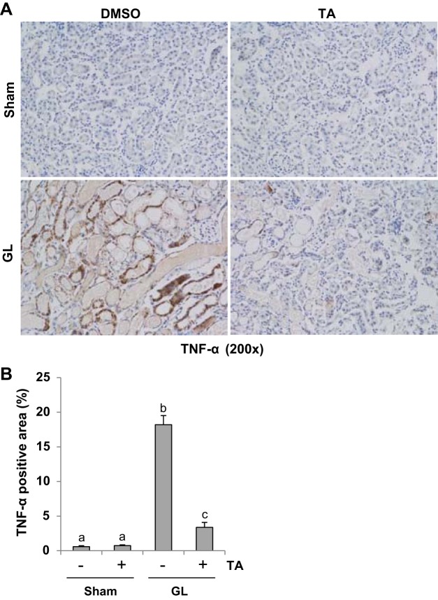
TA significantly reduced expression of TNF-α in GL-induced AKI injured kidneys. A: photomicrographs (×200) illustrating TNF-α-stained sections of kidney tissue on 48 h after GL treatment as indicated. B: TNF-α staining graphic presentation of quantitative data. Data are represented as the means ± SE (n = 6). Means with different superscript letters are significantly different from one another (P < 0.05).

