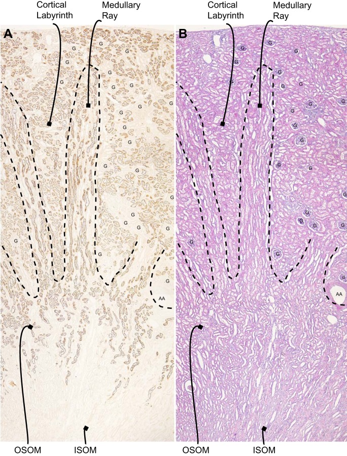
Fig. 2.
Distribution of NaDC1 immunolabel in the human kidney. A: low-power magnification of NaDC1 immunolabel in the human kidney. Immunoreactivity is present in a subset of cells in the cortex in the labyrinth, in the medullary ray in the cortex, and in the outer stripe of the outer medulla (OSOM). No expression is observed in the inner stripe of the outer medulla (ISOM). B: hematoxylin and eosin (H&E) staining of a serial section of the same kidney. G, glomerulus; AA, arcuate artery.

