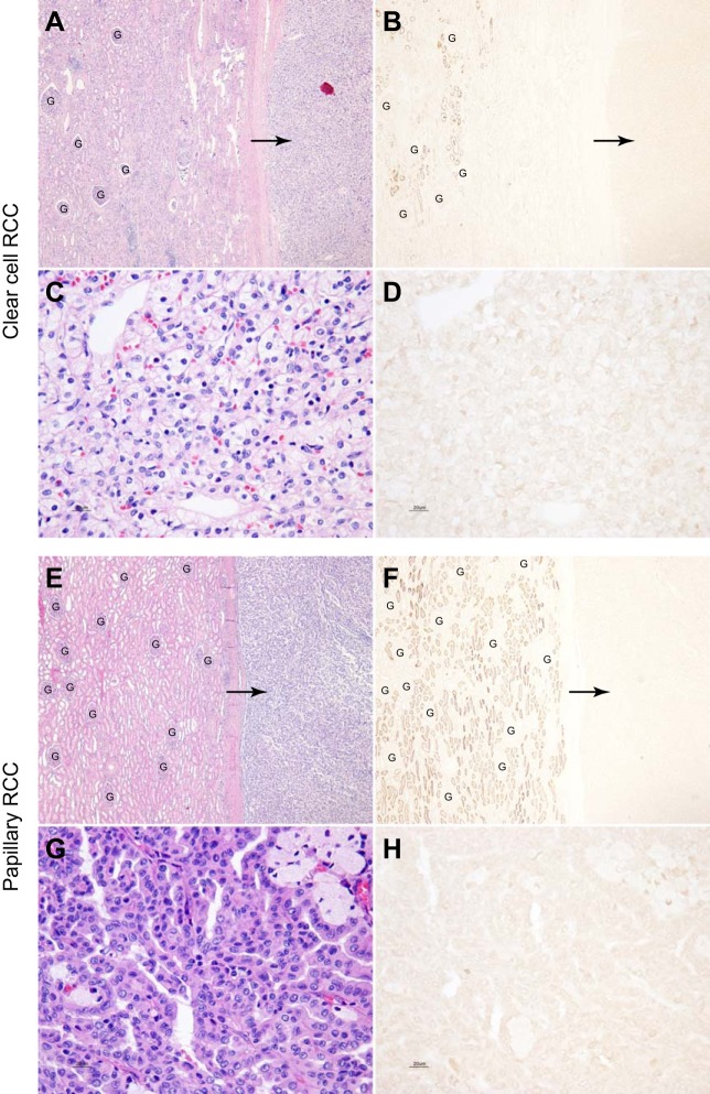
Fig. 5.
NaDC1 expression in the clear cell and papillary renal cell carcinoma. A and B show low-power magnification of hematoxylin and eosin staining of a clear cell renal cell carcinoma (RCC) and adjacent normal renal cortical tissue and NaDC1 immunolabel in a serial section of the same human kidney tissue. No NaDC1 immunolabel is evident in the clear cell carcinoma (arrows). Normal NaDC1 apical immunolabel is present in proximal tubule cells in uninvolved adjacent renal tissue. C and D show high-power magnification of hematoxylin and eosin staining of a clear cell RCC and NaDC1 immunolabel in a serial section of the same clear cell RCC. No detectable NaDC1 immunoreactivity is observed in clear cell RCC. E and F show low-power micrographs of hematoxylin and eosin staining of a papillary RCC and adjacent normal renal cortical tissue and NaDC1 immunolabel in a serial section of the same human kidney tissue. No NaDC1 immunolabel is evident in the papillary RCC (arrows). Normal NaDC1 apical immunolabel is present in proximal tubule cells in uninvolved adjacent renal tissue. G and H show high-power magnification of hematoxylin and eosin staining of a papillary RCC and NaDC1 immunolabel in a serial section of the same a papillary RCC. No detectable NaDC1 immunoreactivity is observed in the papillary RCC. These findings indicate that NaDC1 expression is not detectable in either clear cell or papillary RCC. Glomeruli (“G”) are present in A, B, E, and F. No detectable NaDC1 immunolabel is present in glomeruli.

