Fig. 4.
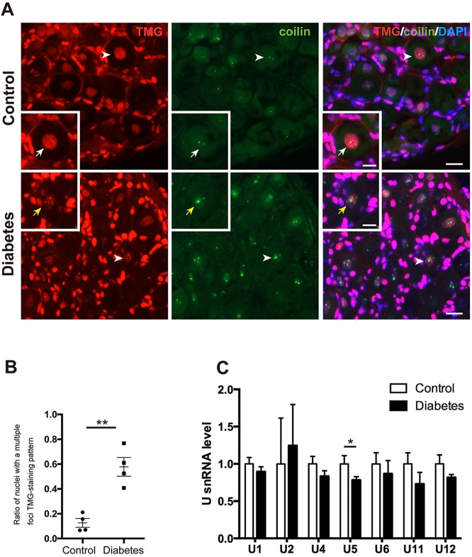
snRNPs form multiple foci with reduction of U5 snRNA and lose their colocalization with CBs in the nuclei of diabetic sensory neurons. (A) TMG staining patterns and relation to CBs (coilin) in diabetic and control mice. snRNPs immunostained for TMG were present homogeneously throughout the nucleoplasm and colocalized with CBs in controls (white arrows), whereas in the diabetic nuclei snRNPs formed multiple foci that did not colocalize with CBs (yellow arrows). Arrowheads indicate sensory neurons magnified in the inset. Scale bar: 20 μm, 10 μm in insets. (B) The ratio of nuclei with snRNPs forming multiple foci was increased in diabetes (n=4) compared with controls (n=4). (C) U snRNA levels in normal (n=6) and diabetic DRGs (n=6). There is a trend towards reduced snRNAs in diabetic sensory neurons. The U5 snRNA level was significantly reduced in diabetic mice compared with controls. *P<0.05, **P<0.01, unpaired one-tailed Student's t-test. Data represented as mean±s.e.m.

