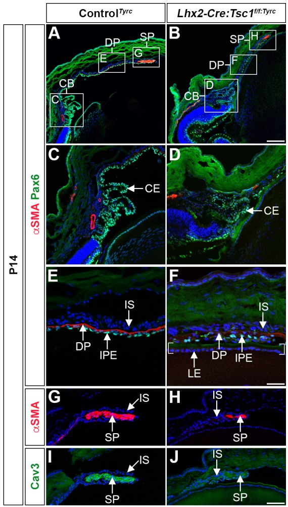
Fig. 3.

Conditional deletion of Tsc1 leads to ciliary body and iris hypoplasia. (A,B) Immunohistochemical analyses of controlTyrc (A) and Lhx2-Cre:Tsc1f/f:Tyrc mice (B) demonstrates atrophic anterior segment structures as defined by Pax6 and αSMA immunoreactivity. (C,D) Immunohistochemical analysis of the CB in controlTyrc (C) and Lhx2-Cre:Tsc1f/f:Tyrc mice (D) shows a disorganised Pax6+ CE and a consequent loss of ciliary processes in mutant animals. (E,F) Immunohistochemical analysis of the medial iris in controlTyrc (E) and Lhx2-Cre:Tsc1f/f:Tyrc mice (F) demonstrates irregularly positioned Pax6+ cells in the IPE of mutant animals. The medial iris of Lhx2-Cre:Tsc1f/f:Tyrc mice also lacks a well-defined DP as demonstrated by a reduced level of αSMA+ immunoreactivity. In this image, the overlying IPE is in close proximity to the LE, which is defined by brackets. (G-J) Immunohistochemical analysis of the proximal iris in controlTyrc (G,I) and Lhx2-Cre:Tsc1f/f:Tyrc mice (H,J) demonstrates that the SP in the mutant animals lacks its well-defined elongated shape and exhibits reduced levels of both αSMA (H) and Cav3 (J). All analyses were performed on mice at P14. Scale bars: 100 µm (A,B), 50 µm (C,D,G,H) and 25 µm (E,F).
