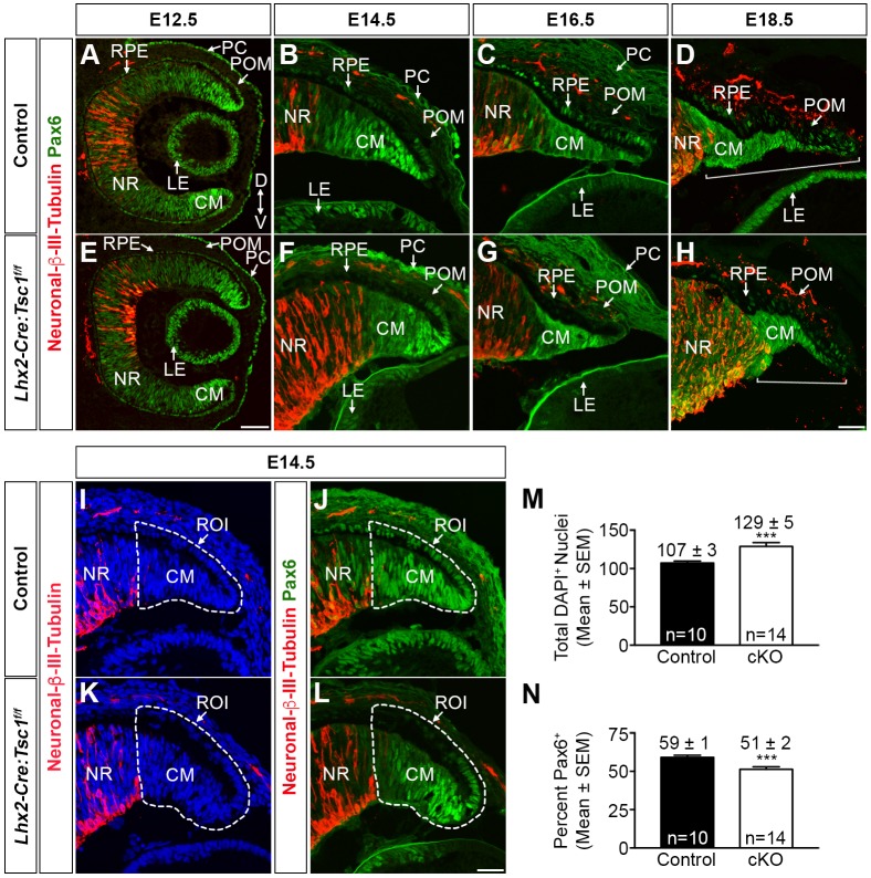
Fig. 5.
Conditional deletion of Tsc1 leads to a reduction in the proportion of Pax6+ cells within the ciliary margin. (A-H) Immunohistochemical analysis of coronal eye sections taken from control (A-D) and Lhx2-Cre:Tsc1f/f (E-H) mice at E12.5 (A,E), E14.5 (B,F), E16.5 (C,G) and E18.5 (D,H) demonstrates that although a comparable centrallow to distalhigh gradient of Pax6 protein distribution is observed at all ages, there is an apparent reduction in the proportion of Pax6+ cells within the CM of mutant animals. A reduction in total CM length is also observed in Lhx2-Cre:Tsc1f/f mice at E18.5 (D,H, brackets) when compared with control animals. CM length is defined as beginning at the distal tip of the CM to the initiation of neuronal β-III-tubulin staining. Note that overlapping neuronal β-III-tubulin and Pax6 immunoreactivity within the NR demarcates developing amacrine and retinal ganglion cells (Ma et al., 2007). (I-N) Quantification of the number of Pax6+ cells in the CM of control and Lhx2-Cre:Tsc1f/f mice at E14.5 using immunohistochemical analysis of Pax6 and neuronal β-III-tubulin. A region of interest (ROI) was drawn around the presumptive CM (I,K, broken line) and the number of DAPI+ cells was then quantified within this ROI. We observed that Lhx2-Cre:Tsc1f/f mice (cKO) had a significant increase in the number of DAPI+ cells within the CM at E14.5 when compared with control littermates (M). The number of Pax6+ cells was then quantified within the ROI (J,L). This Pax6 data was subsequently divided by the DAPI counts to determine the percentage of Pax6+ cells. We observed that Lhx2-Cre:Tsc1f/f mice (cKO) had a significant reduction in the percentage of Pax6+ cells at E14.5 when compared with control littermates (N). All quantification data represent mean±s.e.m. (n=10 eyes for control and n=14 eyes for Lhx2-Cre:Tsc1f/f). ***P≤0.001, calculated using an unpaired two-tailed Student's t-test. Scale bars: 50 µm (A,E); 25 µm (B-D,F-H,I-L).

