Fig. 6.
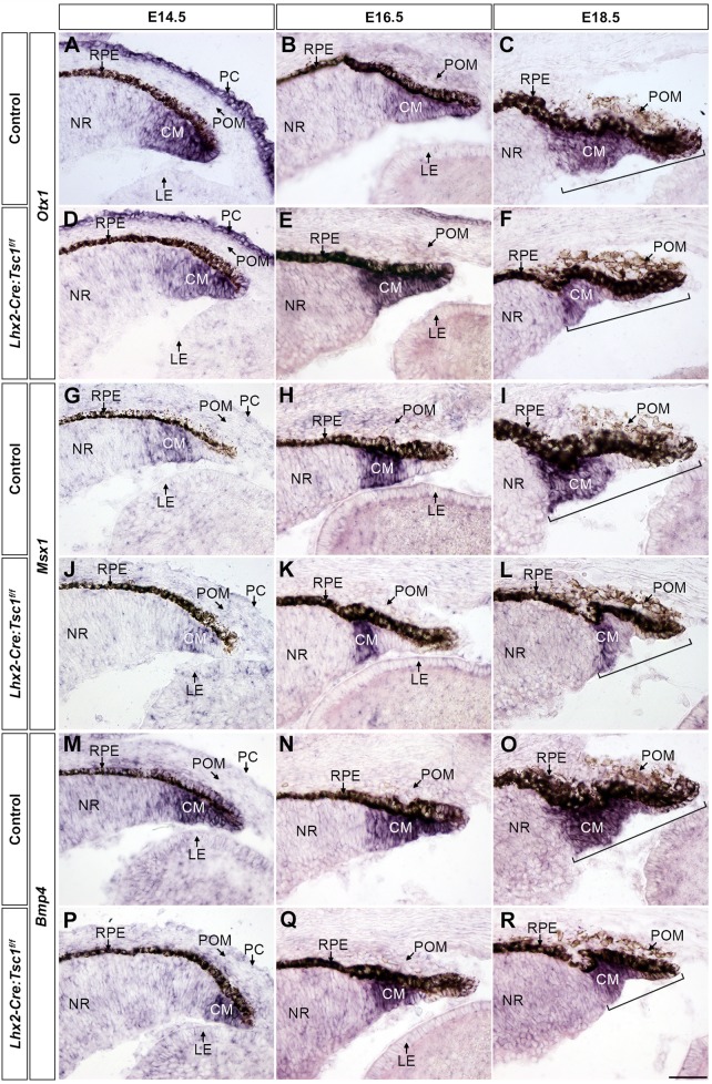
Conditional deletion of Tsc1 disrupts transcriptional programs within the ciliary margin. (A-F) In situ hybridisation analysis of coronal eye sections taken from control (A-C) and Lhx2-Cre:Tsc1f/f (D-F) mice at E14.5 (A,D), E16.5 (B,E) and E18.5 (C,F) demonstrates a comparable expression of Otx1 within the CM at E14.5 (A,D) and E16.5 (B,E). Also note the expression of Otx1 in the overlying PC at E14.5 (A,D). (G-L) In situ hybridisation analysis of coronal eye sections taken from control (G-I) and Lhx2-Cre:Tsc1f/f (J-L) mice at E14.5 (G,J), E16.5 (H,K) and E18.5 (I,L) demonstrates a reduction in the number of Msx1-expressing cells within the CM of mutant animals at all ages analysed. (M-R) In situ hybridisation analysis of coronal eye sections taken from control (M-O) and Lhx2-Cre:Tsc1f/f (P-R) mice at E14.5 (M,P), E16.5 (N,Q) and E18.5 (O,R) shows a reduction in the number of cells that express Bmp4 within the CM of mutant animals at E14.5 (M,P). No difference in Bmp4 expression is seen at E16.5 (N,Q). In addition, a reduction in total CM length at E18.5 is consistently observed in Lhx2-Cre:Tsc1f/f mice when compared with control animals. CM length is defined as beginning from the distal tip of the CM to the proximal part of the Otx1, Msx1 and Bmp4 expression domains (C,F,I,L,O,R, brackets). Scale bar: 50 µm (A-R).

