Fig. 8.

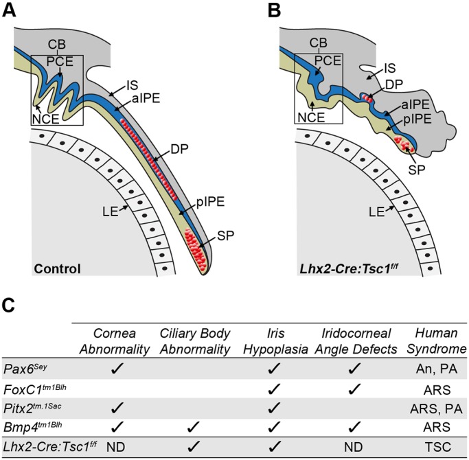
Summary of the anterior segment phenotypes observed in Lhx2-Cre:Tsc1f/f mice and comparison to established mouse models of ASD. (A) Control mice exhibit a CB with well-defined ciliary processes. Iris extension follows the curvature of the lens with the medial region containing the DP and the distal tip containing the SP. (B) Lhx2-Cre:Tsc1f/f mice exhibit hypotrophy of anterior eye structures. While the NCE and PCE of the CB are both present, the overall structure lacks the presence of well-defined ciliary processes. Moreover, the iris fails to extend and presents as a hypotrophic structure with DP and SP atrophy clearly evident. (C) Comparison of anterior segment phenotypes observed in Lhx2-Cre:Tsc1f/f mice with selected phenotypes documented in established mouse models of ASD: Pax6Sey (Hill et al., 1991), Foxc1tm1Blh (Kume et al., 1998), Pitx2tm.1Sac (Gage et al., 1999) and Bmp4tm1Blh (Chang et al., 2001). aIPE, anterior iris pigment epithelium; An, aniridia; ARS, Axenfeld–Rieger syndrome; ND, not determined; PA, Peter's anomaly; pIPE, posterior iris pigment epithelium; TSC, tuberous sclerosis complex.
