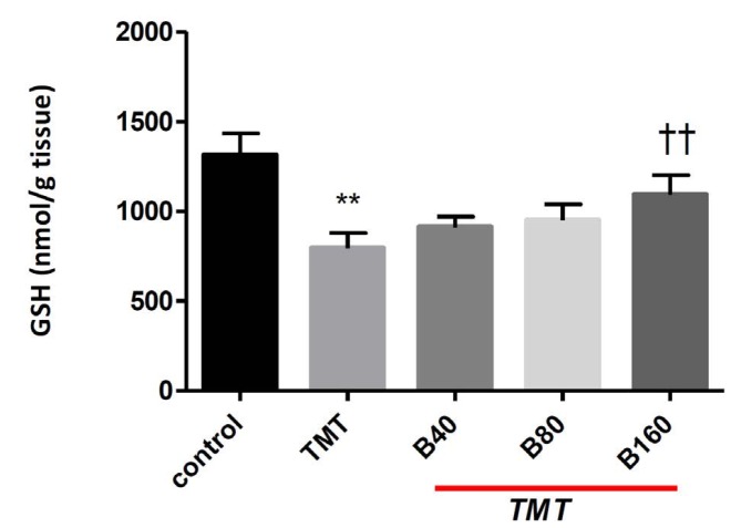
Fig. 3. Effect of BA on GSH content in the cerebral cortex following treatment with TMT. Animals were treated with different doses of BA (40, 80 and 160 mg/ kg) after treatment with TMT at 8 mg/kg). Data are expressed as the means ± SD (n = 8). **P < 0.01 compared with the control group; ††P < 0.01 compared with the TMT-lesioned + saline group.
BA, boswellic acid; GSH, glutathione; TMT, trimethyltin; SD, standard deviation.

