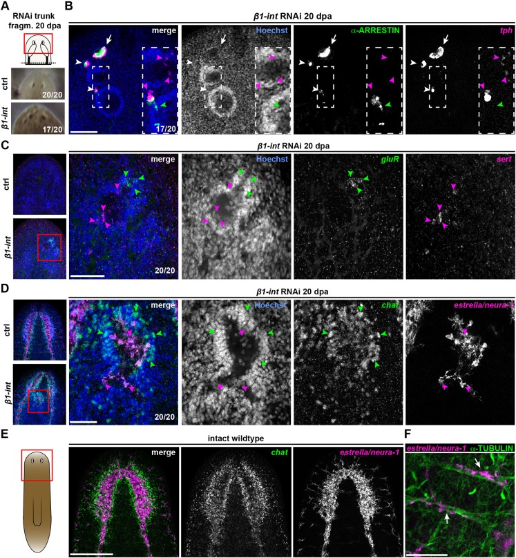
Fig. 6.
Ectospheres are of multineural identity. (A) Live images of anterior regeneration site of ctrl and β1-int RNAi trunk fragments at 20 dpa indicating ectopic eyes in β1-int RNAi animals. (B) Anti-ARRESTIN immunostaining (green; photoreceptor neurons) combined with FISH against tph (magenta) reveals the formation of ectopic eyespots (white arrowheads) with axonal projections towards ectospheres (green arrowheads), next to normal eyespots (white arrows) in β1-int RNAi fragments at 20 dpa. White boxes indicate magnified areas. Magenta arrowheads indicate tph+ cells in ectospheres. (C,D) FISH on β1-int RNAi fragments at 20 dpa against neuronal markers gluR+ (green), sert+ (magenta), chat+ (green) and estrella/neura-1+ (magenta). Magenta or green arrowheads indicate marker+ cells. (E) Double FISH against estrella/neura-1 (magenta) and chat (green) on intact wild-type animals. (F) Anti-α-TUBULIN immunostaining (green) combined with FISH against estrella/neura-1 (magenta). White arrows indicate cells in close proximity to α-TUBULIN+ axon bundles in wild-type animals. Red boxes in schemes illustrate areas of images of A-E. DNA is in blue (Hoechst). Scale bars: 100 µm (B); 50 µm (C,D,F); 250 µm (E).

