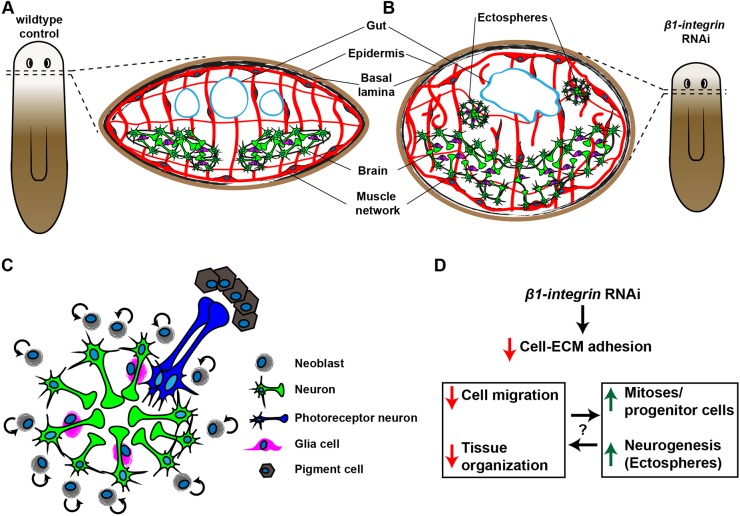
Fig. 7.
The β1-int RNAi phenotype. (A,B) Scheme of tissue organization in regenerating control (A) and β1-int RNAi fragments (B) at 20 dpa. β1-int RNAi planarians revealed severe regeneration defects of gut (blue line), brain (green and magenta) and musculature (red), and ectosphere formation. (C) Ectospheres are composed of neural cells including different neuronal subtypes (green) and glia cells (magenta). Occasionally they contact ectopic eyespots in their proximity [photoreceptor neurons (blue) and pigment cells (brown)]. Ectospheres are polar spheroids with neuronal cell bodies on the outside and axonal projections and glia cells on the inside, displaying a similar organization as the planarian brain. Proliferating neoblasts (gray) in the extracellular environment are likely to account for growth of ectospheres. (D) Summary of the effects of β1-integrin RNAi.

