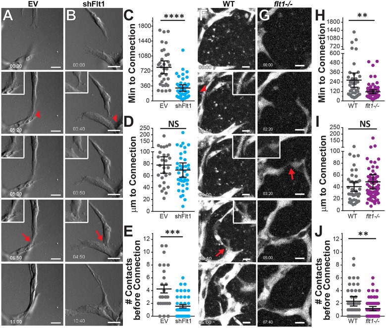
Fig. 3.
Transient contact dynamics during vessel anastomosis are regulated by Flt1. (A,B) Representative time-lapse stills of d3-5 HUVEC sprouts infected with lentivirus encoding (EV) control (A) and shFlt1 (B). Transient contact, red arrowhead; stable connection, red arrow. Insets show higher magnification of vessel interactions. Time, h:min. EV control, n=27 sprouts; shFlt1, n=41 sprouts. (C-E) Quantification of time and distance to connection and the number of contacts made before stable connection for experiments illustrated in A,B. (F,G) Representative time-lapse stills of WT (F) and Flt1−/− (G) ESC-derived vessels at d6 of differentiation. Transient contact, red arrowhead; stable connection, red arrow. Insets show higher magnification of vessel interactions. (H-J) Quantification of time and distance to connection and the number of contacts made before stable connection for experiments illustrated in F,G. WT, n=88 sprouts; Flt1−/−, n=48 sprouts. Two-tailed Student's t-test; **P<0.01, ***P<001, ****P<0.0001; NS, not significant. Error bars, mean±95% CI. Scale bars: 25 µm in A,B; 10 µm in F,G.

