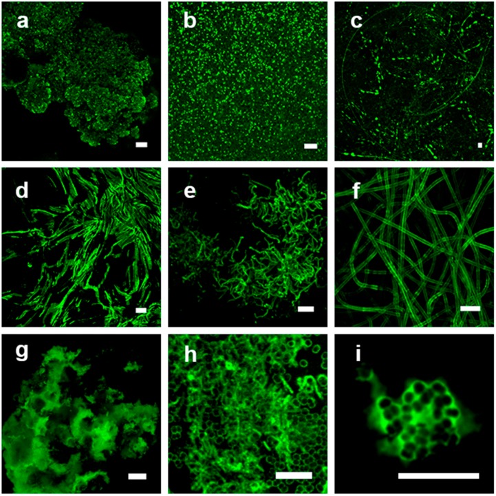
Figure 2.
Confocal laser scanning microscopy of lectin stained microbiological samples representing a variety of binding patterns. The single-channel image data sets are shown as maximum intensity projection. (a) Cauliflower-like glycoconjugate distribution within a bio-aggregate, Metallospaera hakonensis; HMA-FITC; (b) cell surface signal from dense bacterial clusters, Streptococcus mitis; WGA-FITC; (c) glycoconjugate tracks of diatoms on a surface, Craspedostaurus australis; AAL-FITC; (d) slimy matrix in between filamentous microorganisms resulting in a negative staining, Pseudanabaena sp.; PHA-FITC; (e) cell surface of filamentous bacteria, Sphingobium sp.; WGA-FITC; (f) sheath of cyanobacteria filaments, Leptolyngbia sp.; LcH-FITC; (g) bio-aggregate with glycoconjugates showing partly a negative staining in non-binding regions, Ferrovum sp.; WFA-FITC; (h) capsule and rolling tracks of surface associated bacteria, Deinococcus geothermalis; PHA-FITC; (i) matrix signal of a microcolony, Deinococcus geothermalis; HAA-FITC. Scale bar = 10 µm.

