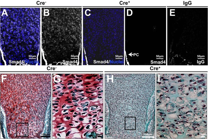
Fig. 2.
Smad4-dependent reduction in growth plate proteoglycan in vivo. (A-E) Immunofluorescence for Smad4 in E18.5 tibial growth plates demonstrate absence of Smad4 in the cartilage of the Col2-Cre+/−;Smad4fl/fl (Cre+) mouse (nuclei: blue, Smad4: gray). (F-I) Safranin O (pink) and Fast Green staining on proximal tibial sections demonstrate a loss of proteoglycan synthesis in the Col2-Cre+/−;Smad4fl/fl growth plate (Cre+; H,I) relative to the Cre− control (F,G). N=3.

