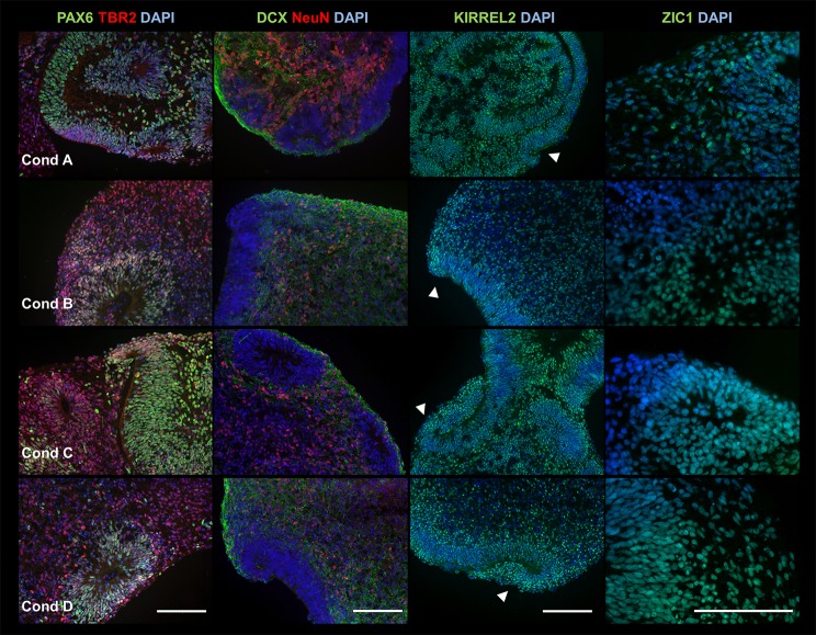
Fig. 3.
FGFs are not required to generate early cortical neuroepithelium. ICC analysis showed that 3D products from medium conditions A-D (rows 1-4, respectively) are all positive for early neural markers PAX6 (green) and TBR2 (red) (first column), neuronal markers DCX (green) and NeuN (red) (second column), and cerebellar neuroepithelium marker KIRREL2 (third column), and granule cell marker ZIC1 (fourth row). Rhombic lip (RL)-like structures are indicated by arrowheads. Experiment was conducted using hESC line H01 and performed three times (n=3) for each condition (A-D). We analyzed 2-3 serial sections per condition, which in total contained the following number of cross sections from a collection of <10 aggregates (one aggregate can consist of several merged structures), condition A (PAX6, 15; TBR2, 15; DCX, 13; NeuN, 13; KIRREL2, 12; ZIC1, 12), condition B (PAX6, 30; TBR2, 30; DCX, 27; NeuN, 27; KIRREL2, 28; ZIC1, 26), condition C (PAX6, 15; TBR2, 15; DCX, 11; NeuN, 11; KIRREL2, 10; ZIC1, 12) and condition D (PAX6, 21; TBR2, 21; DCX, 23; NeuN, 23; KIRREL2, 21; ZIC1, 20). Sample images shown are from hESC line H01. Scale bar: 100 μm.

