Fig. 4.
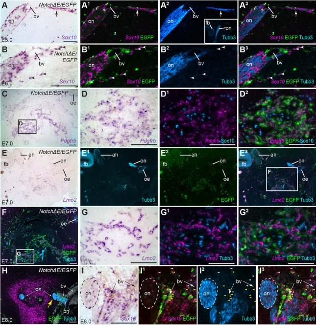
Peripheral axons and glia seem to be attracted to blood vessels containing NotchΔE/EGFP-targeted cells. Parasagittal (A-G2) and coronal (H-I3) sections from embryos in which the cranial ectoderm had been targeted in ovo at E1 with NotchΔE/EGFP, using the Tol2 transposase/‘Tet-on’ electroporation system. Eggs were injected with doxycycline at E4. (A) In an E5 embryo, in situ hybridisation for Sox10 reveals developing OECs on the olfactory nerve. (A1-A3) Same section as A, immunostained for EGFP and Tubb3, with Sox10 shown as a false-colour overlay in A2,A3. A thin nerve branch (arrow) deviates from the olfactory nerve away from the forebrain (for orientation, see low-power inset in A2). The branch-point is near a developing blood vessel, whose wall contains NotchΔE/EGFP-targeted cells. (B-B3) In an E6 embryo, several untargeted Sox10-positive cells (arrowheads), presumably developing OECs, are found isolated in the mesenchyme at some distance from the olfactory nerve, near NotchΔE/EGFP-targeted cells. (C-D2) In an E7 embryo, in situ hybridisation for Pdgfrb followed by immunostaining for EGFP and Sox10 reveals that many NotchΔE/EGFP-targeted cells have formed Pdgfrb-positive perivascular cells, with which many Sox10-positive cells (presumably peripheral glial cells) are associated. This is far from the olfactory nerve: note the location of the olfactory epithelium at the top right. (E-G2) A nearby section of the same E7 embryo, shown at low-power in E-E3 for orientation (note the position of the olfactory epithelium and olfactory nerve towards the top right, and the forebrain and adenohypophysis towards the top left). In situ hybridisation for Lmo2 and immunostaining for EGFP and Tubb3 confirm the presence of peripheral axons (and possibly neurons) close to a large concentration of NotchΔE/EGFP-targeted cells that are associated with Lmo2-positive vascular endothelium. (H) In an E8 embryo (coronal section), the entire olfactory nerve on one side is misplaced laterally (yellow arrow) towards several large blood vessels whose walls contain NotchΔE/EGFP-targeted cells. The displaced olfactory nerve is in contact with another peripheral nerve, and no longer surrounded by cartilage (identified by immunostaining with an anti-Sox9 antibody that also cross-reacts with other SoxE family members), unlike the olfactory nerve on the other side. (I-I3) In a nearby section of the same E8 embryo, in situ hybridisation for Sox10 and immunostaining for EGFP and Tubb3 show that some Sox10-positive OECs – both untargeted (black/white arrowheads) and NotchΔE/EGFP-targeted (yellow arrowheads) – are found at a distance from axons, associated instead with blood vessels whose walls contain NotchΔE/EGFP-targeted cells. ah, adenohypophysis; bv, blood vessel; EGFP, enhanced GFP; fb, forebrain; oe, olfactory epithelium; on, olfactory nerve; pn, peripheral nerve. Scale bars: 100 µm.

