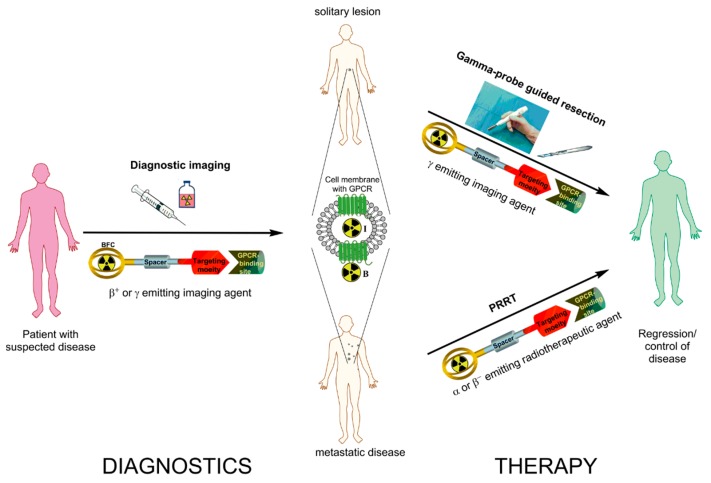
Figure 1.
Schematic representation of radiotheranostics using radiolabeled peptides targeting G-protein coupled receptors (GPCR). The radiopharmaceutical consists of the targeting moiety (peptide), a bifunctional chelator (BFC) forming a stable complex with the radionuclide, and often a spacer in between the two units. Radiotheranostics involves diagnostic (left panel) and therapeutic (right panel) components. In targeting of GPCRs (somatostatin receptor (sstr), glucagon-like peptide-1 receptor (GLP-1R), cholecystokinin 2 receptor (CCK2R) or glucose-dependent insulinotropic polypeptide receptor (GIP-R); middle panel) the radiotheranostic agent would bind to GPCR and either internalize into the cell, when utilizing an agonist (I) or stay on the surface of the cell, when utilizing an antagonist (B). In the first step diagnostic imaging using β+ or γ emitting imaging agent to assess the spread of disease is conducted. On the therapeutic stage, in the given example, the patient is subjected either to the gamma-probe guided resection or Peptide Receptor Radionuclide Therapy (PRRT). In radioguided surgery pre-administration of corresponding γ emitting imaging agent gives real-time information to the surgeon and guides the localization of the GPCR rich tumor site intraoperatively. In the case of a metastatic disease α or β− emitting radiotherapeutic agent is administered to target the GPCR rich lesions internally (PRRT).

