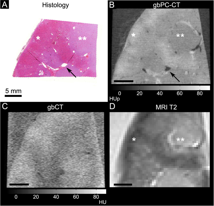
Figure 1. Normal human kidney sample imaged with phase-contrast CT, grating-based attenuation-based CT, T2-w magnetic resonance imaging and histologic slice (coronal slice).
Good visual agreement between the histology ((A); HE-staining) and grating-based phase-contrast CT ((B); gbPC-CT) showed a higher soft-tissue contrast and a clear discrimination of renal vessels (arrow) and between the cortex (*) with higher and the medulla (**) with lower phase-contrast signal with good comparison to T2-w magnetic resonance imaging (MRI) (D). Imaging with grating-based attenuation-based CT (gbCT) from the same setup (C) had an obvious lower soft-tissue contrast.

