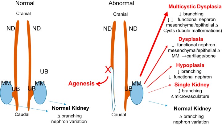
Fig. 2.
Macroview of normal and abnormal kidney development. Normal development of the kidneys involve bilateral extension of the pronephric/mesonephric duct (ND) proceeding in a cranial-to-caudal direction. Ultimately, the ureteric bud (UB) grows out from ND, invades the metanephric mesenchyme (MM), and undergoes a series of branching (as described in detail in Fig. 1). Slight changes in the timing, expression, and function of genes/proteins likely account for the observed natural variation in branching and nephron number. For conditions of abnormal development, such as failure of a kidney to develop and/or other congenital defects including ipsilateral urogenital tissues, such as vas deferens, seminal vesicle and epididymis, or uterine horn (Mullerian duct) result from a truncation of the ND. Likewise, more profound alterations in timing, expression, and function of kidney genes/proteins could lead to a variety of congenital kidney defects.

