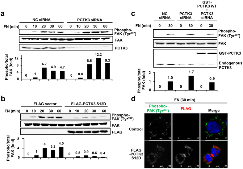
Figure 5. PCTK3 suppresses fibronectin-mediated FAK activation during early cell adhesion.
(a,b) PCTK3 siRNA (a) or FLAG-tagged PCTK3 S12D (b) transfected HEK293T cells were plated on fibronectin (FN)-coated dishes for indicated times. The cell lysates were subjected to immunoblot analysis with anti-phospho-FAK (Tyr-397), anti-FAK, anti-FLAG, and anti-PCTK3 antibodies. (c) HEK293T cells were transfected with NC or PCTK3 siRNA. After 24 hours, cells were transfected with empty GST vector or GST-tagged PCTK3 wild type for 24 hours. Cells were plated on FN-coated dishes for 30 minutes. The cell lysates were subjected to immunoblot analysis with anti-phospho-FAK (Tyr-397), anti-FAK, and anti-PCTK3 antibodies. The levels of phosphorylated FAK (Tyr-397) were normalized to the levels of total FAK. The phosphorylation of FAK (Tyr-397) in fibronectin-stimulated NC siRNA-transfected cells were taken as 1. (d) FLAG-PCTK3 S12D-transfected HEK293T cells were plated on FN-coated dishes for 30 minutes. Then, cells were fixed and immunostained with anti-phospho-FAK (Tyr-397) and anti-FLAG antibodies. Fluorescence for phospho-FAK (Tyr-397) and FLAG is shown in green and red, respectively. Hoechst nuclear staining is represented in blue.

