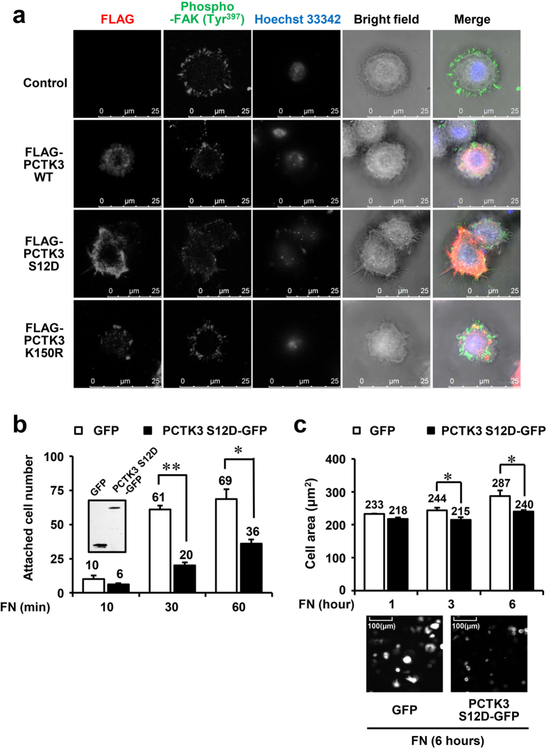
Figure 6. PCTK3 leads to filopodia formation and a reduction in cell adhesion in HeLa cells.
(a) PCTK3 WT, S12D, or K150R-expressing HeLa cells were plated on fibronectin-coated coverslips for 30 minutes. The cells then were fixed and stained with anti-FLAG and anti-phospho-FAK (Tyr-397) antibodies. Fluorescence for FLAG and phosphor-FAK (Tyr-397) are shown in red and green respectively. Hoechst nuclear staining is represented in blue. The images were captured by using a 63× objective lens. (b,c) HeLa cells transfected with GFP or PCTK3 S12D-GFP were detached and suspended. Then, cells were plated at a density of 5 × 10⁴ cells per well on fibronectin-coated 12 well plates and incubated for indicated times. Cells were washed with PBS and fixed. The cell area and attached number of GFP expressing cells were counted using a 40× objective lens of IN Cell Analyzer 6000. Results are expressed as means ± S.E. from six independent fields. Statistical significance was determined by Student’s t-test. *p < 0.05 and **p < 0.01. Inset: Immunoblot analysis with anti-GFP antibody.

