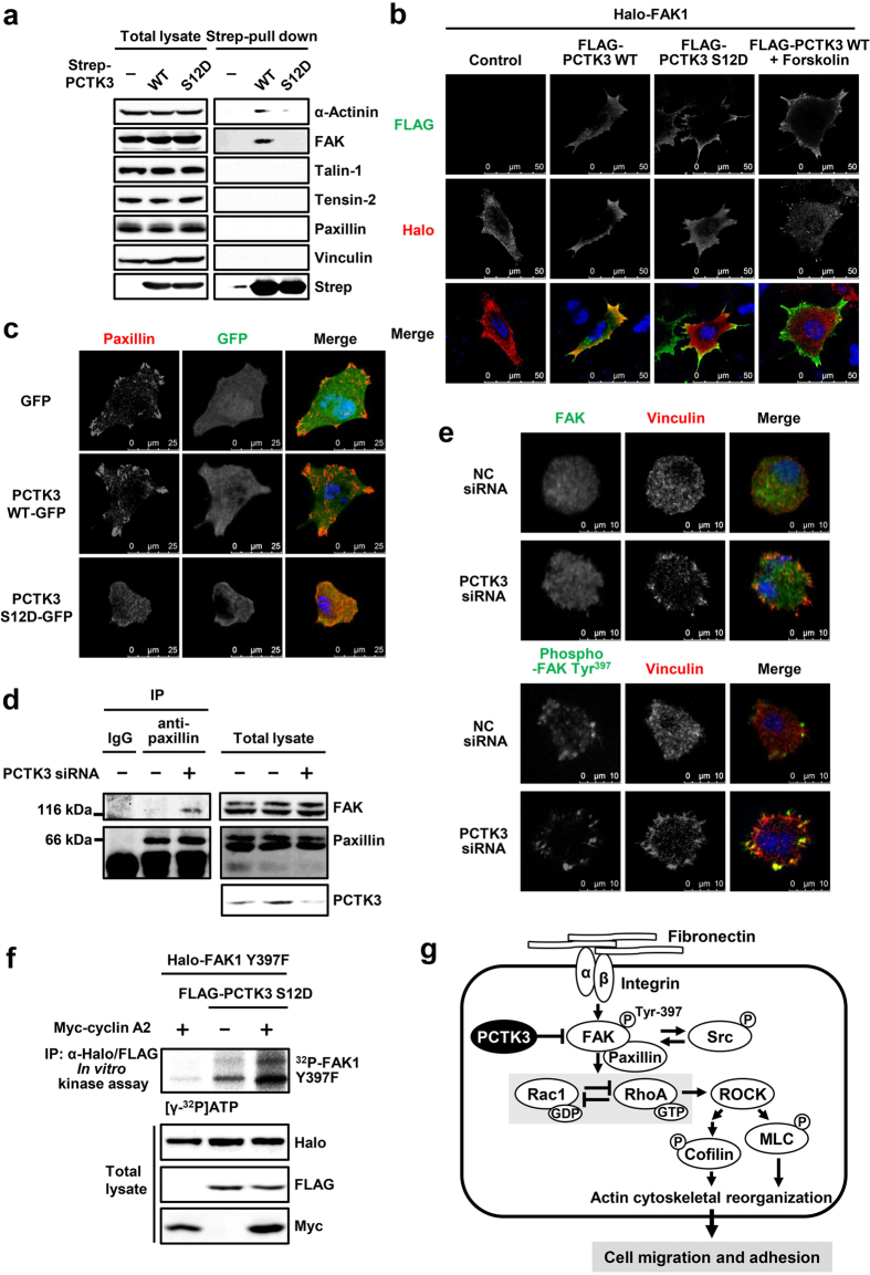
Figure 7. PCTK3 activation induces the dissociation from focal adhesion.
(a) HEK293T cells were transfected with Strep-tagged PCTK3 wild type (WT) or S12D mutant. Cell lysates were subjected to Strep-pull down assay and immunoblot analysis with anti-α-actinin, anti-FAK, anti-talin-1, anti-tensin-2, anti-paxillin, anti-vinculin, and anti-Strep antibodies. (b) HeLa cells were transfected with Halo-tagged FAK1 together with FLAG-tagged PCTK3 WT or S12D. After treatment with 20 μM forskolin or DMSO for 30 minutes, cells were fixed and immunostained with anti-Halo and anti-FLAG antibodies. Fluorescence for Halo and FLAG are shown in red and green, respectively. Hoechst nuclear staining is represented in blue. The images were obtained using a 63× objective lens. (c) GFP-, PCTK3 WT-GFP-, or PCTK3 S12D-GFP-expressing HeLa cells were plated on fibronectin-coated coverslips. After 3 hours, cells were fixed and immunostained with anti-paxillin antibody. Fluorescence for GFP and paxillin are shown in green and red, respectively. Hoechst nuclear staining is represented in blue. (d) The lysates of PCTK3-knockdown cells were subjected to immunoprecipitation with anti-paxillin antibody or control IgG. The immunoprecipitates or lysates were analyzed by immunoblotting using anti-FAK, anti-paxillin, and anti-PCTK3 antibodies. (e) PCTK3 siRNA-transfected HEK293T cells were plated on fibronectin-coated dishes for 30 minutes. Then, cells were fixed and stained with anti-FAK, anti-phospho-FAK (Tyr-397), and anti-vinculin antibodies. Fluorescence for FAK and phospho-FAK (Tyr-397) are shown in green. Fluorescence for vinculin is shown in red. Hoechst nuclear staining is represented in blue. (f) FLAG-PCTK3 S12D was expressed with Halo-FAK Y397F in the presence or absence of Myc-cylin A2 in HEK293T cells. After immunoprecipitation using anti-FLAG and anti-Halo antibodies, the precipitated samples were incubated in a kinase buffer containing [γ − 32P]ATP for 30 min, and subjected to SDS-PAGE, after which the gel was analyzed by bioimaging analyzer. The expression levels of FLAG-tagged, Halo-tagged, and Myc-tagged proteins were confirmed by immunoblotting with anti-FLAG, anti-Halo, and anti-Myc antibodies, respectively. (g) A model of the PCTK3/FAK signaling pathway.

