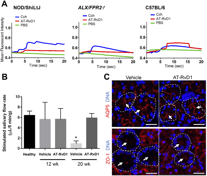
Figure 1. Treatment with AT-RvD1 prior to disease onset maintains healthy saliva flow rates and restores tissue integrity in female NOD/ShiLtJ mice.
(A) Submandibular gland (SMG) cell clusters were obtained from 20 wk NOD/ShiLtJ, ALX/FPR2−/− and C57BL/6 mice, mounted on coverglass and stimulated with AT-RvD1 (100 ng/mL), carbachol (Cch; positive control; 100 μM), or PBS (negative control) to measure changes in intracellular free calcium concentrations as described in the Material and Methods. Results shown are from a representative experiment from three or more determinations. (B) Salivary flow rates were calculated using female mice treated with either a vehicle control (8.9% EtOH in saline) or AT-RvD1 (0.1 mg/kg) at 4, 12 and 20 wk. Results from N = 3 mice were used per condition and data are expressed as mean ± SD, with *P < 0.05 indicating a significant difference from controls. (C) SMG from female NOD/ShiLtJ mice treated with AT-RvD1 or a vehicle control, were harvested, formalin-fixed, paraffin-embedded, and sectioned. Then, aquaporin 5 (AQP5) and zonula occludens-1 (ZO-1) were localized using confocal microscopy. TO-PRO-3 Iodide was used as a nucleic acid stain (blue). White arrows indicate luminal structures. Note that both AQP5 and ZO-1 appear disorganized in control mice. Representative fluorescent images from N = 3 are shown. Scale bars represent 50 µm.

