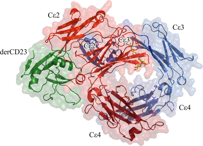
Figure 1. Structure of the 1:1 derCD23/IgE-Fc complex.

The derCD23 (green Cα traces with surfaces) binds to one heavy chain of IgE-Fc (red and blue) contacting the Cε3 and Cε4 domains. The Cε2 domains are asymmetrically bent back onto one Cε3 domain and make some additional contacts with derCD23. The carbohydrate is shown in all-atom representation (red and yellow, without surfaces) and can be seen behind the Cε3 domain.
