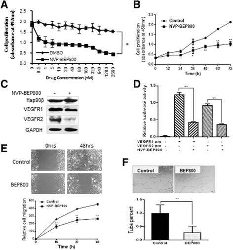
Fig. 5.

NVP-BEP800 inhibited endothelial cell proliferation, migration, invasion, and tube formation. a Incubation with different concentrations of NVP-BEP800 for 48 h inhibited HUVEC cell proliferation. b Growth curve of HUVEC cells incubated with 2 μM NVP-BEP800, as measured by SRB assay. c Western blot analysis showed that treatment with 2 μM NVP-BEP800 down-regulated VEGFR1 and VEGFR2 expression in HUVEC cells. d HUVEC cells were transiently transfected with VEGFR1- or VEGFR2- dependent reporter gene plasmids and treated with 2 μM NVP-BEP800. After 24 h, the cells were lysed and luciferase activity was measured. e The wound healing ability of HUVEC cells was assessed after 48 h of NVP-BEP800 treatment. DMSO treatment was used as the standardized control for quantification. f In vitro assay for vascular mimicry of HUVEC cells treated with NVP-BEP800 for 48 h in three-dimensional culture. NVP-BEP800 inhibited cord formation of HUVEC cells. *P < 0.05, **P < 0.01
