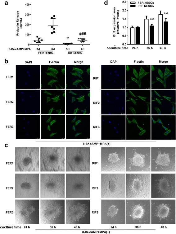
Fig. 1.

Impaired decidualization of hESCs isolated from endometrium of patients with RIF. a hESCs isolated from both RIF patients and fertile controls were treated with 0.5 mM 8-Br-cAMP and 1 μM MPA as indicated for an additional 3 days or 6 days. dPRL released into the medium was detected by ELFA. ** P < 0.01 compared with fertile hESCs after treatment for 3 d; ### P < 0.001 compared with fertile hESCs after treatment for 6 d, repeated measures data ANOVA. b Fluorescein isothiocyanate-labeled phalloidin was used to label actin filaments, and immunofluorescence was adopted to analyze the morphological transformation of hESCs from both fertile controls (n = 6) and RIF patients (n = 6) after treatment with 0.5 mM 8-Br-cAMP and 1 μM MPA for 3 days. c Attachment of BLSs to hESC monolayers isolated from both RIF patients (n = 6) and fertile controls (n = 6) were assessed following treatment with 0.5 mM 8-Br-cAMP and 1 μM MPA as indicated for an additional 72 h. Several spheroids were transferred to confluent hESC monolayers from different women. Cells were imaged after 24 h, 36 h, and 48 h. d The area of BLS expansion on the FER hESCs and RIF hESCs were detected at different coculture time points and is presented relative to that of the 24 h time point (area was set to 1). *** P < 0.001 compared with FER hESCs group at the same time point, repeated measures data ANOVA
