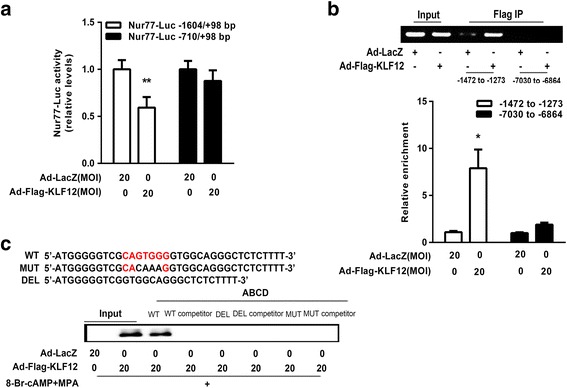
Fig. 4.

KLF12 directly repressed Nur77 transcription. a hESCs were infected with the indicated adenoviruses for 24 h and then transfected with Nur77-Luc (−1604/+98 bp) or deleted Nur77-Luc (−710/+98 bp) (300 ng/well). After 48 h, luciferase assays were performed, and the data were plotted after normalization to Renilla luciferase activity. ** P < 0.05 compared with Ad-LacZ alone, Student’s t-test. b ChIP-PCR amplification using primers against the human Nur77 promoter region (top). PCR products were separated by agarose gel electrophoresis. Quantitative ChIP analysis was performed by real-time PCR. The results are shown as fold changes relative to LacZ (after normalization to the input DNA, bottom). Input (non-precipitated) chromatin was utilized as a positive control in these analyses. * P < 0.05 compared with Ad-LacZ alone, Student’s t-test. c ABCD assays were performed using biotinylated or non-biotinylated (competitor) double-stranded Nur77 wild-type (WT), conserved element-deleted (DEL) and conserved element-mutated (MUT) oligonucleotides with whole-cell extracts from hESCs
