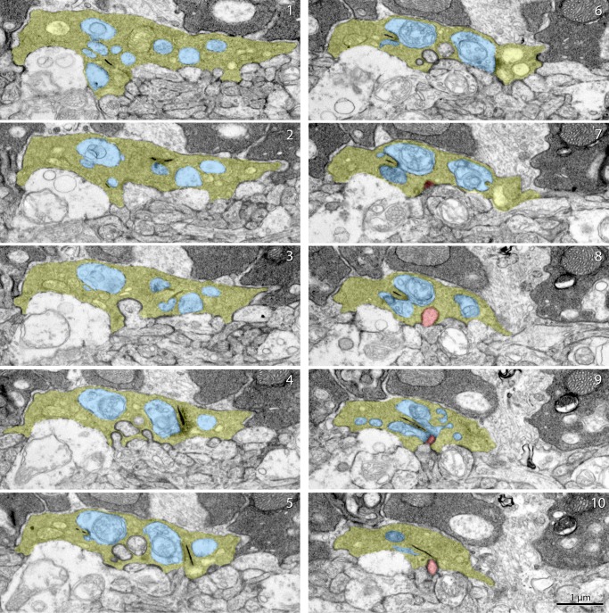
Figure 5.
Serial ultrathin section images of a cone pedicle in an Lrit3nob6/nob6 retina. Serial electron microscopy images of a cone pedicle in an Lrit3nob6/nob6 retina. A gallery shows images of every two sections. Cone pedicle is shown in yellow, invaginating dendrite of cone ON-BC is shown in red, and horizontal cell processes are shown in blue. Scale bar: 1 μm.

