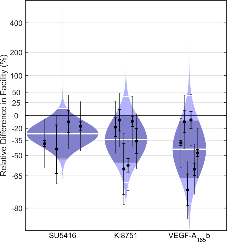
Figure 5.
Antagonists to VEGFR-2 decrease outflow facility in enucleated mouse eyes. Cello plots showing the relative difference in C between contralateral eyes of C57BL/6 mice perfused with or without 3 μM SU5416, 1 nM Ki8751, or 0.5 μg/ml human VEGF-A165b. Ki8751 (P = 0.04, n = 6, weighted t-test) and VEGF-A165b (P = 0.03, n = 6) reduced facility by 34% (CI: −56, −2%) and 44% (CI: −66, −8%) on average, respectively, while SU5416 reduced facility on average by 27% (CI: −54, 14%) but did not achieve significance (P = 0.10, n = 4). Data points represent the relative facility difference of a treated eye with respect to its contralateral untreated eye for individual pairs. The thick white lines represent the geometric mean of the relative difference for each group. The remaining symbols are as defined in Figure 2.

