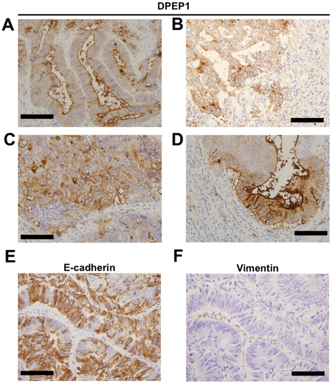
Figure 2.
Immunohistochemical staining of DPEP1 in CRC. Representative image of DPEP1 staining of (A-D) tumorous tissue samples, (E) E-cadherin and (F) Vimentin. (A) Apical, (B) cytoplasmic, (C) circumference and (D) mixed staining patterns for DPEP1 in CRC. (E) Positive staining pattern for E-cadherin in CRC. (F) Negative staining pattern for Vimentin in CRC. Scale bar=100 µm. DPEP1, dipeptidase 1; CRC, colorectal cancer; E-cadherin, epithelial cadherin.

