FIGURE 2.
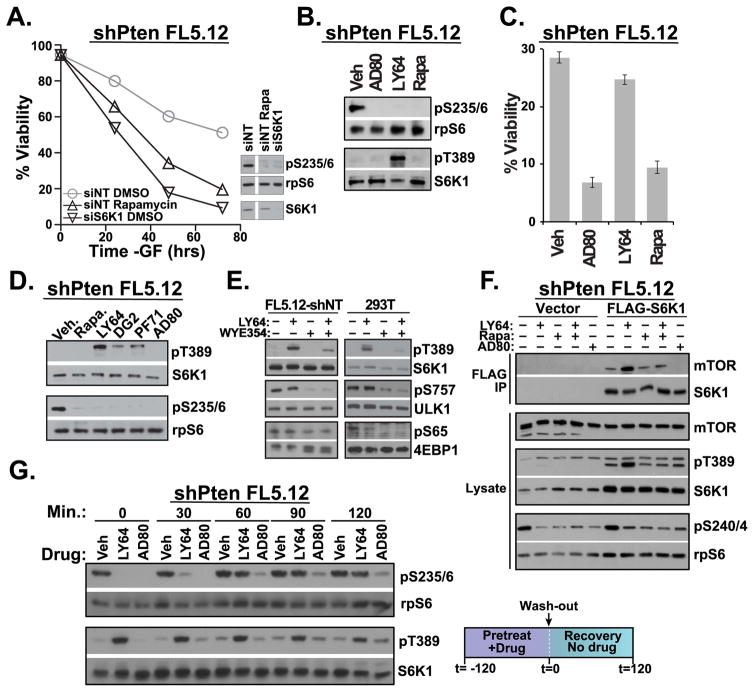
S6K1 inhibitor pathway dynamics. (a) IL-3-dependent FL5.12 cells that had been stably transduced with shPTEN were cultured in full medium lacking only IL-3. Transfection of siS6K1 siRNA, or treatment with 20 nM rapamycin induced apoptosis in PTEN-deficient cells. n=3. (b) PTEN-deficient FL5.12 cells cultured –IL-3 for 1 hour in the presence of 4 μM AD80, 1 μM LY-2779964 (LY64) or 20 nM rapamycin (Rapa) revealed reduced rpS6 phosphorylation yet substantial differences in S6K1 T389 phosphorylation. n=2. (c) Viability of cells treated as in (b) for 72 hours. (d) 1 μM LY-2779964, 10 μM DG2, and 10 μM PF-4708671 share an ability to increase pT389 when added to PTEN-deficient FL5.12 cell cultures for 2 hours. 4 μM AD80 avoids the induction of pT389. n=2. (e) shNT FL5.12 cells or serum-starved 293T cells were incubated with 1 μM LY-2779964 or the mTOR ATP-competitive inhibitor WYE354 (1 μM) for 30 minutes. n=4, FL5.12-shNT; n=2, 293T. (f) PTEN-deficient FL5.12 cells transduced with either vector or FLAG-S6K1 were cultured in the indicated inhibitors for 3 hours, prior to lysis and FLAG-IP. n=3. (g) PTEN-deficient FL5.12 cells were preincubated with vehicle control or 1 μM LY-2779964 or 10 μM AD80 for 2 hours. Cells were then washed and phosphorylation kinetics determined by immunoblot. n=3. See also Figure S3.

