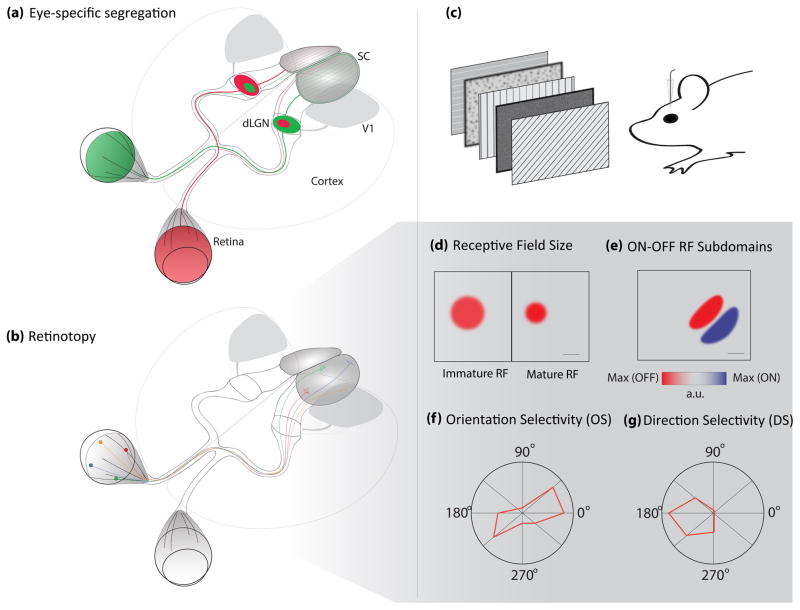
Figure 2.
Examples of the major receptive field properties referred to in this review. (a–b) Large scale circuit refinement properties. (a) Eye-specific lamination is here depicted as the projections from each retina in a different color to their targets in the LGN and SC. In higher mammals, thalamocortical projections also tessellate V1 with ocular dominance columns. (b) Projections from four retinal positions are shown with their corresponding targets in the SC where their retinotopic positions are preserved. Retinotopy is also present in dLGN, V1 and extrastriatal visual areas, but is not shown for clarity. (c) Schematic depicting common methodology for recording receptive fields in mice while presenting various visual stimuli. (d) Example of an RF measured from a single ON responsive cell before activity-dependent refinement (left) and that same cell after refinement (right). Scale bar indicates 2 visual degrees (see reference 34). (e) Schematic of the ON and OFF responses of a single neuron and their corresponding RF positions. In this example the neuron responds to an elongated ON (red) region in space (where an increment in light best produces a response), that is close but not overlapping with an OFF (blue) field (where a decrement in light best produces a response) Scale bar indicates 20 visual degrees (see reference 81). (f) Example of an orientation selective (OS) neuron that responds preferentially to gratings in two opposite directions, thus non-selective for direction but rather for the orientation of the moving bars. Each spoke on the rose plot represents a direction of motion of drifting gratings presented to the mouse (see 2c). The amplitude along each direction represents the relative strength of firing for a neuron to a given direction. (g) Lastly, a direction-selective (DS) response example where this neuron only responds to leftward movement. Both OS and DS are most highly tuned in V1 in all species studied, but also occur in subcortical regions, at a seemingly higher frequency in rodents and rabbits than other mammals with higher visual acuity. Likewise, OS in subcortical regions of mice is less sharp than OS in the cortex.

