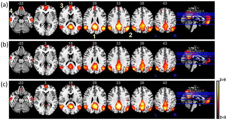
Fig. 1.

The DMN for (a) healthy controls (HC), (b) PDAR (akinetic/rigidity-predominant) and (c) PDT (tremor-predominant) subjects. The DMN as shown above is consistent with previously reported results and highlights the possible differences in activity between PDT/PDAR subtypes and HCs. Images are in radiological orientation. 1) posterior cingulate cortex (PCC), extending dorsally into the precuneus; 2) bilateral inferior parietal cortex (left and right IPC); 3) medial prefrontal cortex (mPFC)/anterior cingulate cortex and 4) medial/lateral temporal lobe (MTL).
