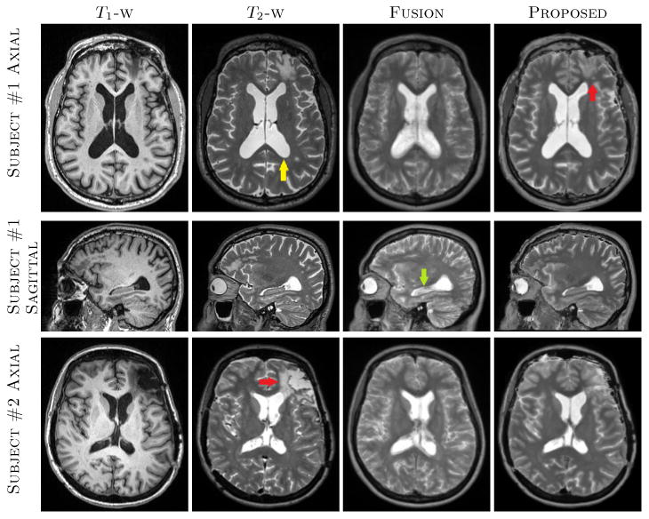
Fig. 1.

The top two rows show axial and sagittal views of a patient from HighRes dataset, where T1-w, T2-w, Fusion [14], and proposed synthesis results are shown. There is a lesion in the frontal lobe (LowRes arrow) which was not synthesized in Fusion. Also ventricles and cortex are fuzzier (yellow arrow) as well as hippocampus (LowRes arrow) has CSF-like intensities in Fusion based T2. The bottom row shows another subject from the same dataset where the lesion on the left frontal lobe (LowRes arrow) is not well synthesized in either synthetic T2s. Our method generally produced sharper features in the cortex and anatomically correct intensities near the hippocampus.
