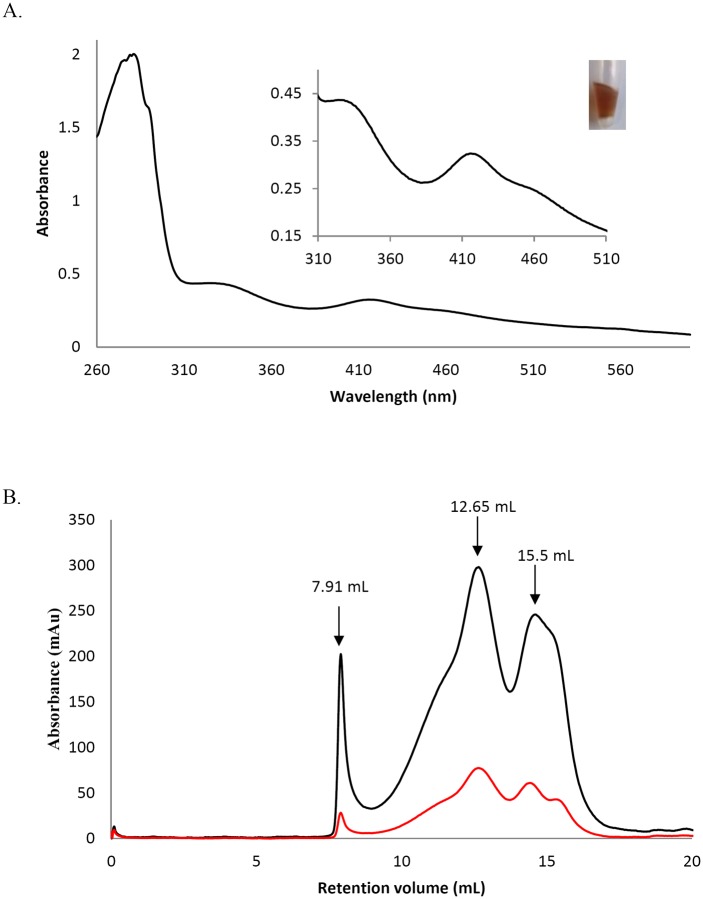
Fig 4. The recombinant PDI-A binds an Fe-S cluster.
A. UV-visible absorption spectrum between 260 and 600 nm of anaerobically purified PDI-A. The blow-up shows absorbance bands between 310 and 510 nm characteristics of a [Fe2-S2] cluster and a picture of a tube containing the protein in its holoform. B. Analytical size-exclusion chromatography performed on a Sephadex S75 10–300 column using 100 μg and a 30mM Tris-HCl pH 8.0, 200 mM NaCl buffer. The black curve corresponds to the absorbance at 280 nm and the red curve at 420 nm.

