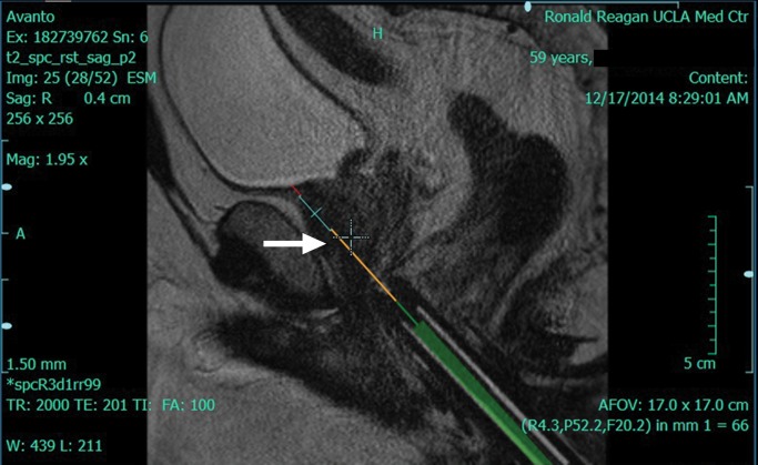
Figure 2c:
DynaCAD workstation (InVivo) images show that the workstation is capable of offering visualization of the tumor during biopsy planning. (a) The lesion is localized (arrow) on an axial T2-weighted image. (b) The software provides target coordinates, which are adjusted on the dial (arrowheads). (c) Target confirmation (arrow) is shown.

