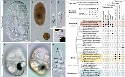
Fig. 1. Diversity and independent origins of extrusomes.

(A to E) Nematocysts in the dinoflagellate P. kofoidii, including a live whole cell (A) and a cell that was preserved in Lugol’s iodide solution while capturing a prey cell of A. tamarense (B). (C) Enhanced contrast image shows the defensive trichocysts deployed by A. tamarense (arrows) in response to attack by P. kofoidii. (D and E) Isolated nematocysts from P. kofoidii, seen as unfired (D) and discharged (E). (F to H) Nematocysts in the dinoflagellate Nematodinium sp., which have an eye-like ocelloid (F). A battery of nematocysts is visible in the live cell (G) and remains intact after cell lysis (H). (I) Genomic distribution of known extrusome proteins (vertical labels) across eukaryotes based on best reciprocal Basic Local Alignment Search Tool (BLAST) hits (black squares), which is a common predictor of protein homology. Taxa are listed within an established phylogenetic framework (38, 39).
