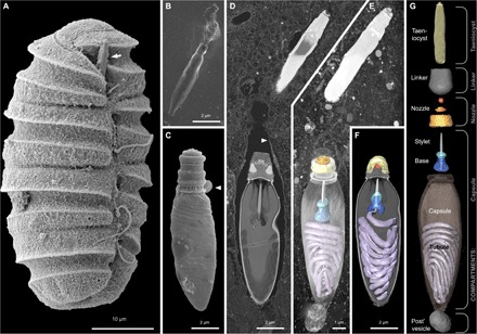
Fig. 2. Reconstruction of the nematocysts in the dinoflagellate P. kofoidii.

(A) SEM micrograph of a cell of P. kofoidii, including an armed taeniocyst (arrow) in the apical region that first contacts prey. (B) SEM micrograph of an isolated taeniocyst that has discharged its amorphous contents. (C) SEM micrograph of an isolated nematocyst that has become arrested very early in discharge; arrowhead, operculum. (D) FIB-SEM section of taeniocyst and nematocyst enclosed by a membranous chute (arrowhead) and (E) maximum intensity projection of the same region seen slightly from above and below (F). (G) Virtual dissection of the nematocyst-taeniocyst complex. Brackets indicate the membrane-bound compartments in which those components are grouped during early development. Later in development, compartments fuse to form the chute.
