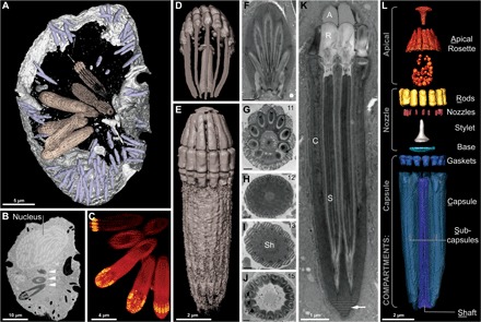
Fig. 4. Reconstruction of the nematocysts in Nematodinium sp.

(A to E) Images derived from single-cell FIB-SEM. (A) Partial reconstruction of a cell with nematocysts shown in beige and mucocysts shown in purple. (B) Longitudinal FIB-SEM section through a cell showing the nucleus and nematocysts (arrowheads). (C) 3D maximum intensity projection showing a battery of nematocysts. (D) 3D tilted reconstruction of a nematocyst. (E) Longitudinal reconstruction of a nematocyst. (F to K) Single-cell TEM micrographs of a nematocyst in oblique section (F), cross section (G to J), and longitudinal section (K) showing the striated material (arrow) and variable symmetries across different nematocysts, with the number of subcapsules printed in the upper right corner. (L) A virtual dissection of the nematocyst components. Brackets indicate the membrane-bound compartments bounding the components indicated. (F) to (J) scale bar, 500 nm.
