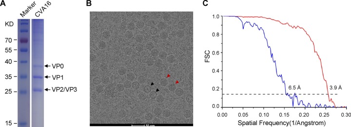
FIG 1.
SDS-PAGE analysis, cryo-EM image, and resolution evaluation of CVA16 viral particles. (A) SDS-PAGE and Coomassie blue staining of purified BPL-inactivated CVA16. The lanes for the inactivated CVA16 sample and the protein ladder marker on the same gel were aligned for molecular mass estimation. (B) Representative cryo-EM micrograph of CVA16. The red and black arrowheads indicate mature virion and empty capsid, respectively. Scale bar, 50 nm. (C) Resolution evaluation of the cryo-EM maps of the BPL-inactivated CVA16 particles by Fourier shell correlation (FSC) at a 0.143 criterion. Red and blue curves represent the FSCs of the mature virion and the empty capsid, respectively.

