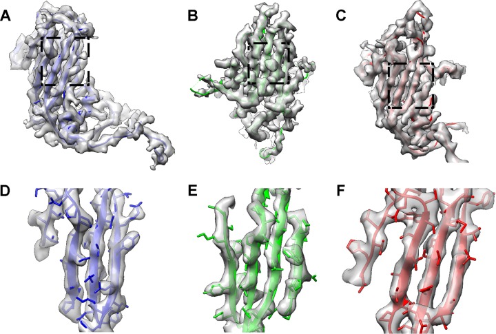
FIG 3.
Fitting of the 135S-like crystal structure into the cryo-EM map of the BPL-inactivated mature virion. (A to C) The crystal structure of the CVA16 135S-like particle (PDB 4JGY) fits well into the corresponding density map. VP1, VP2, and VP3 are shown in blue, green, and red, respectively. The same color scheme is followed throughout. (D to F) Expanded views of panels A to C (indicated by the dashed rectangles), illustrating the high-resolution structure features, including separation of beta strands as well as the majority of bulky side chains.

