
FIG 5.
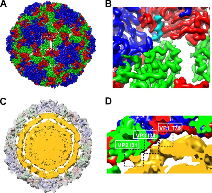
Features of the cryo-EM structure of the BPL-inactivated CVA16 mature virion. (A) Externalization of the VP1 N terminus from the base of canyon. The density for residues 62 to 70 of VP1 is rendered in cyan. Densities for other parts of VP1, VP2, and VP3 are rendered in blue, green, and red, respectively. (B) Expanded view of panel A. (C and D) Cross section of BPL-inactivated CVA16 virion structure that was low-pass filtered to 10-Å resolution. The capsid is rendered in transparent gray with the fitted atomic model. The RNA shell and core are rendered in yellow. (D) Expanded view of panel C. Three contacts between the capsid and the RNA shell are indicated by dashed rectangles, and the residues that contributed to the contacts are labeled.