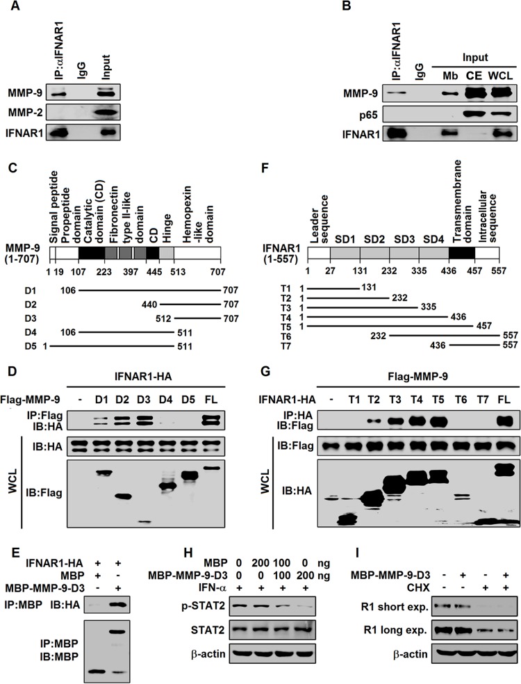
FIG 7.
MMP-9 interacts with IFNAR1 at the cell surface. (A) Differentiated THP-1 cells (5 × 10⁶) were lysed, and cell lysates were immunoprecipitated with anti-IFNAR1 or IgG. The immunoprecipitates and WCLs were analyzed by Western blotting with anti-MMP-9, anti-MMP-2, and anti-IFNAR1. (B) Mbs of differentiated THP-1 cells (5 × 10⁶) were immunoprecipitated with anti-IFNAR1 or IgG. The immunoprecipitates, Mbs, CEs, and WCLs were analyzed by Western blotting with anti-p65, anti-MMP-9, and anti-IFNAR1. (C) Schematic diagram of wild-type MMP-9 (MMP-9) and truncated mutants (MMP-9-D1 to -D5). (D) HEK293T cells were cotransfected with plasmid pCAGGS-IFNAR1 expressing HA-tagged IFNAR1 and plasmid p3×FLAG-CMV-14-MMP-9 expressing Flag-tagged MMP-9 (FL) or plasmids p3×FLAG-CMV-14-MMP-9-D1 to -D5 expressing Flag-tagged MMP-9-D1 to -D5. Cell lysates were immunoprecipitated with anti-Flag. The immunoprecipitates and WCLs were analyzed by Western blotting with the indicated antibodies. IB, immunoblotting. (E) MBP pulldown assay with lysates from IFNAR1-HA-expressing HEK293T cells and MBP or MBP-MMP-9-D3 purified from E. coli cells. After pulldown, the precipitates were analyzed by Western blotting with anti-HA and anti-MBP. (F) Schematic diagram of wild-type IFNAR1 (IFNAR1) and truncated mutants (IFNAR1-T1 to -T7). (G) HEK293T cells were cotransfected with plasmid p3×FLAG-CMV-14-MMP-9 and plasmid pCAGGS-IFNAR1 expressing full-length HA-tagged IFNAR1 or plasmids pCAGGS-IFNAR1-T1 to -T7 expressing HA-tagged truncated IFNAR1 (IFNAR1-T1 to -T7). Cells were lysed, and cell lysates were immunoprecipitated with anti-HA antibody. The immunoprecipitates and WCLs were analyzed by Western blotting with anti-Flag and anti-HA. (H) HepG2 cells were treated with MBP or MBP–MMP-9-D3 for 3 h and treated with rhIFN-α for 30 min. p-STAT2, STAT2, and β-actin levels were determined by Western blotting. (I) HepG2 cells were treated with MBP or MBP–MMP-9-D3 for 12 h and treated with CHX (50 μg/ml) for 4 h. IFNAR1 and β-actin levels were determined by Western blotting.

