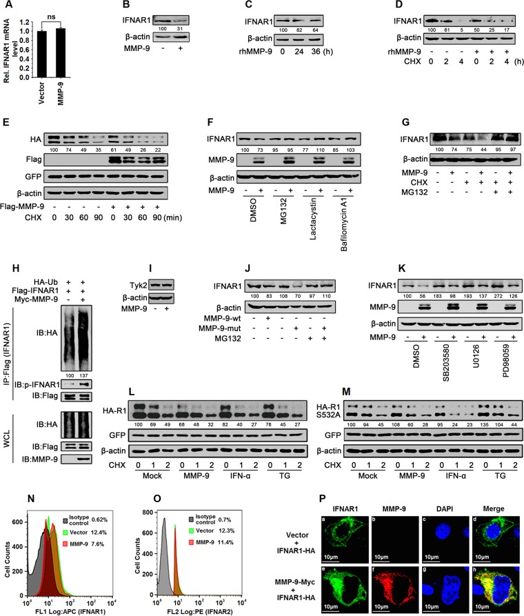
FIG 8.
MMP-9 promotes the phosphorylation, ubiquitination, degradation, and subcellular distribution of IFNAR1. (A and B) HepG2 cells were transfected with pCMV-Tag2B-MMP-9 for 48 h. Cells were harvested. IFNAR1 mRNAs were analyzed by qPCR (A), and IFNAR1 and β-actin proteins were detected by Western blotting (B). (C) HepG2 cells were treated with rhMMP-9 (50 ng/ml) for different times. Cells were harvested, and IFNAR1 and β-actin were detected by Western blotting. (D) HepG2 cells were treated with rhMMP-9 (50 ng/ml) for 12 h and treated with CHX (50 μg/ml) for the indicated times. Cells were harvested, and IFNAR1 and β-actin were detected by Western blotting. (E) HEK293T cells were cotransfected with pCAGGS-IFNAR1 and pCMV-Tag2B-MMP-9 for 48 h and treated with CHX (50 μg/ml) for the indicated times. The proteins were analyzed by Western blotting using the corresponding antibodies. (F) HepG2 cells were transfected with pCMV-Tag2B-MMP-9 for 42 h and treated with proteasome inhibitors (MG132 and lactacystin) and a lysosome inhibitor (bafilomycin A1) for 6 h. Cells were harvested, and IFNAR1, MMP-9, and β-actin were detected by Western blot analyses. (G) HepG2 cells were transfected with pCMV-Tag2B-MMP-9 for 42 h and treated with MG132 (20 μM) for 2 h and with CHX (50 μg/ml) for 4 h. Cells were harvested, and IFNAR1 and β-actin were detected by Western blot analyses. (H) HEK293T cells were cotransfected with pCDNA3.1-HA-Ub, pCDNA3.1-3×Flag-IFNAR1, and pcDNA3.1-Myc or pcDNA3.1-Myc-MMP-9 and treated with MG132 (20 μM) for 9 h. Cell lysates were denatured and subjected to IP with anti-Flag. The immunoprecipitates and WCLs were analyzed by Western blotting with the indicated antibodies. (I) HepG2 cells were transfected with pCMV-Tag2B or pCMV-Tag2B-MMP-9 for 48 h. TYK2 and β-actin protein levels expressed in the treated cells were determined by Western blot analyses using the corresponding antibodies. (J) HepG2 cells were transfected with pCMV-Tag2B-MMP-9-wt or pCMV-Tag2B-MMP-9-mut for 42 h and treated with MG132 (20 μM) for 6 h. Cells were harvested, and IFNAR1 and β-actin were detected by Western blot analyses. (K) HepG2 cells were transfected with pCMV-Tag2B-MMP-9 for 42 h and treated with the signaling component inhibitors SB203580 (p38 MAPK inhibitor), U0126 (ERK1/2 inhibitor), and PD98059 (MEK inhibitor) for 6 h. Cells were harvested, and levels of IFNAR1, MMP-9, and β-actin proteins were detected by Western blot analyses. (L and M) HEK293T cells were cotransfected with pCAGGS-IFNAR1 (L) or pCAGGS-IFNAR1-S532A (M) and pCMV-Tag2B-MMP-9 for 33 h and treated with rhIFN-α or TG (1 μM) for 1 h and with CHX (50 μg/ml) for the indicated times. Levels of IFNAR1-HA, IFNAR1-S532A–HA, GFP, and β-actin proteins were determined by Western blotting. (N and O) HepG2 cells were transfected with pCMV-Tag2B-MMP-9 for 48 h and then harvested. IFNAR1 protein (N) and IFNAR2 protein (O) levels were analyzed by flow cytometry. APC, allophycocyanin; PE, phycoerythrin. (P) HepG2 cells were cotransfected with pCAGGS-IFNAR1 and pcDNA3.1-Myc-MMP-9 for 24 h. Cells were immunostained with anti-Myc and anti-HA antibodies, and the nucleus was stained by DAPI and analyzed by confocal microscopy. Results show means ± standard deviations. ns, nonsignificant.

