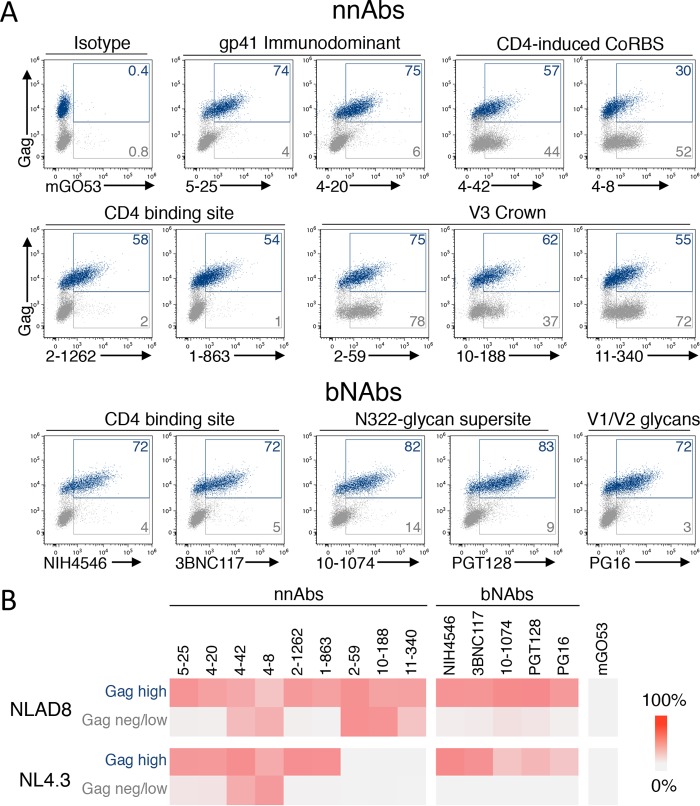
FIG 1.
Antibody binding at the surface of cells infected with two HIV-1 reference strains. (A) CEM-NKR cells infected with HIV-1 (NLAD8) were incubated with 15 μg/ml of anti-Env monoclonal antibodies. The levels of antibody bound on infected (Gag-high) and bystander (Gag-low and Gag-negative) cells were then evaluated by flow cytometry. Dead cells were excluded based on morphological criteria using side and forward scatters. A representative dot plot of each indicated antibody is presented. (B) CEM-NKR cells infected with HIV-1 (NLAD8 or NL4.3) were stained with the indicated antibodies, and the percentage of antibody-positive cells was measured by flow cytometry. The heat map represents the mean percentage of Ab+ cells in infected (Gag-high) or bystander (Gag-negative/low) cells obtained from 3 independent experiments.

