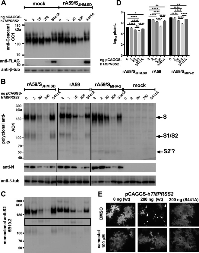
FIG 3.
TMPRSS2 overexpression decreases productive MHV infection. (A) TMPRSS2 decreases CEACAM1a protein levels. HEK-293T cells cotransfected with pCAGGS-mCeacam1a-4L and pCAGGS-hTMPRSS2-FLAG or pCAGGS-hTMPRSS2-S441A-FLAG were infected with rA59/SJHM.SD-EGFP and lysed for immunoblotting at 18 hpi. (B) TMPRSS2 decreases cell-associated MHV protein levels. HEK-293T cells cotransfected with pTK-mCeacam1a-4L and pCAGGS-hTMPRSS2-FLAG or pCAGGS-hTMPRSS2-S441A-FLAG were infected as indicated and lysed for immunoblotting 18 hpi. Goat polyclonal anti-S antibody AO4 was used to detect the S protein and mouse anti-N monoclonal antibody 1-16-1 to detect N protein. The vertical lines indicate boundaries between nonadjacent lanes (rA59/SJHM.SD-EGFP and rA59-EGFP were run on the same gel, but their positions were exchanged for consistency with other panels; rA59/SMHV-2-EGFP and the mock-infected cells were run in parallel on a separate gel). (C) TMPRSS2 cleavage of S may be nonproductive. Probing the lysates shown in panel C with anti-S2 MAb 5B19.2, previously mapped to the fusion peptide, detected an ∼150-kDa fragment (black box) inconsistent with S2′ cleavage. (D) TMPRSS2 decreases MHV titer. HEK-293T cells were cotransfected with pTK-mCeacam1a-4L and 200 ng of pCAGGS-hTMPRSS2-FLAG or pCAGGS-hTMPRSS2-S441A-FLAG and infected with the indicated viruses; cell supernatants were collected at 18 hpi and titers determined. Both active and inactive TMPRSS2 significantly decreased the MHV titer (using 2-way ANOVA with Dunnett's multiple comparisons of each TMPRSS2 level with the 0-ng control within each virus, P < 0.0001 for the effect of the virus, P < 0.0001 for the effect of TMPRSS2 transfection, and P < 0.0045 for the interaction; *, P < 0.05; **, P < 0.01; ***, P < 0.001; and ****, P < 0.0001 for the multiple comparisons). Data are representative of 2 independent experiments performed in triplicate. (E) TMPRSS2 activity decreases syncytium size. HEK-293β5 cells were cotransfected with pTK-mCeacam1a-4L and pCAGGS-hTMPRSS2-FLAG or pCAGGS-hTMPRSS2-S441A-FLAG and infected as described for panels B to D, treated at 2 hpi with DMSO or camostat as indicated (final DMSO concentration of 0.1% for all treatments), and fixed for microscopy at 18 hpi.

