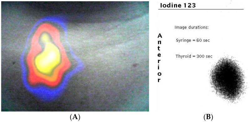
Figure 9.
(A) Combined gamma and optical image of a patient’s thyroid during clinical investigation. The uptake of 123I is clearly seen in the right lobe of the patient’s thyroid (left side of image); (B) the standard clinical image taken by a large field of view gamma camera in the nuclear medicine clinic.

