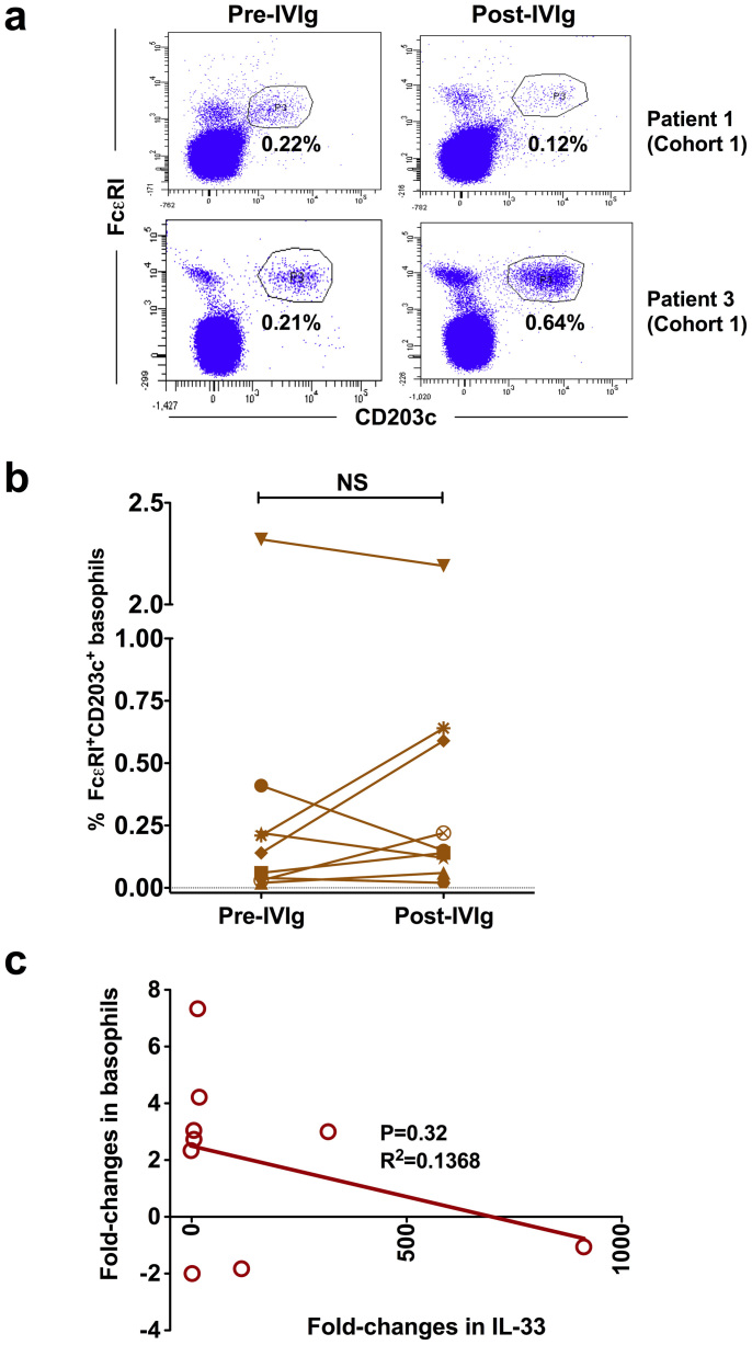
Figure 2. Changes in the proportion of circulating basophils of autoimmune patients following IVIg therapy.
Heparinized blood samples were obtained from cohort 1 patients with inflammatory myopathies before (Pre-IVIg) and 2–3 days after initiation of IVIg therapy (Post-IVIg). (a) Representative dot-plots showing basophils from cohort 1 patients gated positive for FcεRI and CD203c (b) Modulation of circulating basophils following IVIg therapy (n = 9). Basophils were analyzed in the whole blood by flow cytometry. The statistical significance as determined by two-tailed Student-t-test is indicated, where NS, non-significant. (c) The correlation between fold-changes in IL-33 and basophils following IVIg therapy.

